— VIENNA, VA • MINUTES FROM TYSONS CORNER

Specialists in Medical Aesthetics and Hormone Optimization in Northern Virginia

Botox, dermal fillers, PDO thread lifts, laser skin renewal and PRP hair restoration delivered by Dr. Sadiya Naseem, DNP, FNP-BC, a national trainer of injectors across the country.

Tag: ultrasound and filler

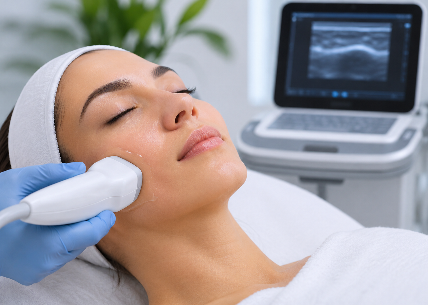
Facial ultrasound examination showing a practitioner scanning a patient's cheek to assess underlying facial tissues and structures.

Facial ultrasound is a non-invasive imaging technique that uses high-frequency sound waves to examine structures beneath the skin. It helps practitioners visualise blood vessels, muscles, fat compartments, and existing filler. This technology supports better anatomical assessment before injectable treatments. Consequently, practitioners can plan more precise procedures and reduce avoidable complications.

Personalised aesthetic care can now come with added convenience. At Sadiya Clinic, Dr. Sadiya Naseem uses ultrasound assessment to support informed treatment decisions and natural-looking results. The clinic also provides concierge services within 25 miles of its Vienna location. These services include Botox, Xeomin, and most fillers. Minimum charges apply depending on the number of clients. Schedule a consultation today to discuss your aesthetic goals and explore suitable treatment options.

How Does Diagnostic Ultrasound Work Beneath the Skin?

Diagnostic facial ultrasound uses high-frequency sound waves to create images of structures beneath your skin. A handheld probe sends sound waves into facial tissues. These waves return as echoes when they meet different structures. The device converts these signals into images. This allows practitioners to examine muscles, fat compartments, connective tissues, and blood vessels.

A 2025 research paper published in Ultrasound in Medicine & Biology [1], highlights the role of diagnostic imaging in aesthetic dermatology. It explains how ultrasound helps examine facial tissues and identify important anatomical structures. Additionally, Doppler imaging supports blood vessel assessment. These methods help practitioners plan injectable treatments with better anatomical awareness. 

How Does Facial Ultrasound Make Injectables Safer?

Facial ultrasound makes injectables safer by helping practitioners identify blood vessels and examine tissue layers before treatment. It provides real-time images of underlying structures. This allows injectors to assess anatomical variations and choose suitable placement depths. Consequently, imaging helps reduce avoidable risks during cosmetic procedures, although it cannot prevent every possible complication.

Here are the main ways ultrasound supports safer injectable treatments:

  • Identifying Blood Vessels: Doppler imaging helps locate arteries and veins near planned injection sites. Practitioners can adjust their approach to avoid identified vessels.
  • Checking Tissue Depth: Ultrasound shows different facial layers beneath the skin. This helps injectors select suitable placement depths for specific products.
  • Detecting Existing Filler: Imaging reveals certain previous filler deposits. Therefore, practitioners can assess their position before adding more products.
  • Supporting Complication Management: Ultrasound helps locate deposits associated with swelling or suspected vascular problems. It can also guide hyaluronidase towards targeted hyaluronic acid filler.
  • Improving Injection Accuracy: Real-time imaging helps practitioners monitor needle or cannula positioning. This provides additional control during selected procedures.

Although facial ultrasound improves anatomical awareness, it does not guarantee complication-free treatments. Results still depend on practitioner training, product selection, and appropriate injection techniques. Furthermore, qualified injectors must understand facial anatomy and recognise warning signs. They should also maintain emergency protocols. Combining these measures with imaging helps support informed decisions and reduce avoidable treatment risks.

Which Treatments Can Use Facial Ultrasound?

Facial ultrasound can support several aesthetic treatments, including dermal fillers, Botox, Sculptra, Radiesse, and Kybella. It also helps practitioners assess previous injections and guide filler dissolving procedures. Each application serves a different purpose. However, imaging requirements depend on the product, treatment area, and individual anatomy.

A 2024 review published in Seminars in Ultrasound, CT and MRI [2], explores ultrasound applications in aesthetic medicine. It covers filler and non-filler procedures, including their imaging requirements. Additionally, the study explains how ultrasound helps practitioners examine facial anatomy and guide injections. These applications support more informed treatment planning and complication assessment. 
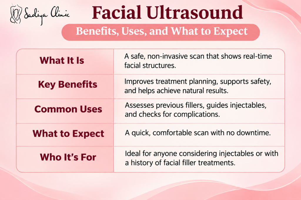
Facial ultrasound explaining its benefits, common aesthetic uses, appointment expectations, and suitable patients.

Who Benefits Most From Ultrasound Guided Injections?

Ultrasound-guided injections can benefit patients who need detailed facial assessment before aesthetic treatments. They are particularly useful for people with previous fillers, uneven results, or concerns about complications. Additionally, individuals considering injections in complex facial areas may benefit from vascular mapping. However, suitability depends on personal anatomy, treatment goals, and practitioner recommendations.

Patients With Previous Filler Treatments

If you have received fillers before, ultrasound can help locate existing deposits. This information helps practitioners assess their position and avoid unnecessary product placement during future procedures.

Individuals Experiencing Filler Migration

Unwanted fullness, swelling, or irregular contours may indicate filler migration. Ultrasound helps examine affected areas and identify deposits that require further assessment or corrective treatment.

Patients Considering High-Risk Treatment Areas

The forehead, temples, and nose contain complex blood vessel networks. Imaging helps practitioners map these structures before injections and select more appropriate treatment approaches.

Individuals Seeking Filler Dissolving

If you want to remove unwanted hyaluronic acid filler, ultrasound can locate specific deposits. This allows practitioners to guide hyaluronidase towards targeted areas more precisely.

Patients With Previous Injection Complications

Persistent lumps, swelling, or suspected vascular problems require careful assessment. Ultrasound provides additional information about underlying tissues and helps practitioners plan appropriate management.

A 2023 review published in Diagnostics [3], highlights ultrasound’s role in personalised aesthetic treatments. It explains how imaging helps assess previous fillers, detect migration, and examine tissue changes. Additionally, practitioners can use this technology to guide targeted hyaluronidase placement. These applications support individual treatment planning and more informed clinical decisions. 

Book Your Ultrasound Guided Consultation at Sadiya Clinic

Previous filler complications, uneven results, or unexpected swelling can make you hesitant about further cosmetic procedures. You might worry about hidden product deposits or accidental vascular injuries. Without proper imaging, understanding your facial anatomy becomes challenging. You deserve personalised care that addresses these concerns before another injectable treatment.

At Sadiya Clinic, facial ultrasound helps practitioners examine underlying structures and make informed treatment decisions. This technology supports the identification of existing filler and nearby blood vessels. It also helps guide precise injection techniques and reduce avoidable risks. Contact us today at sadiya@sadiyaclinic.com to arrange your consultation and explore suitable aesthetic options.

FAQs

Is Facial Ultrasound Painful?

No, facial ultrasound is generally painless and comfortable. The practitioner applies gel and gently moves a handheld probe across your skin. You may feel slight pressure during scanning. However, the process requires no needles or incisions. It uses sound waves instead of radiation, making it a gentle imaging method.

Is Facial Ultrasound the Same as HIFU or Ultherapy?

No, facial ultrasound is different from HIFU and Ultherapy. Diagnostic ultrasound creates images of facial structures beneath the skin. In contrast, HIFU and Ultherapy deliver focused energy into deeper tissues. This heat stimulates collagen production and supports skin tightening. Therefore, both technologies serve different aesthetic purposes.

Can Ultrasound Show Old Filler in My Face?

Yes, facial ultrasound can identify many existing filler deposits beneath your skin. High-frequency imaging helps practitioners locate their position, depth, and distribution. It can also reveal filler that has moved beyond its original placement. However, visibility depends on the product type, tissue changes, and time since injection.

Does Ultrasound Guided Filler Cost More?

Yes, ultrasound-guided filler may cost more than traditional injections, depending on the clinic. Additional equipment, scanning time, and practitioner expertise can influence pricing. However, fees vary between providers and treatment areas. Ask your clinic whether imaging is included in the procedure cost or charged separately before booking your appointment.

Where Can You Get Ultrasound Guided Injections in Vienna, VA?

You can find ultrasound-guided injections at specialised aesthetic practices in Vienna, VA. Look for qualified medical injectors with facial ultrasound training and experience in vascular mapping. Before booking, ask about their imaging equipment, injection techniques, and complication management protocols. This helps you choose a provider focused on treatment precision and patient safety.

About the Author

Dr. Aimen Arij, Pharm.D.

Dr. Aimen Arij, Pharm.D., is a licensed Clinical Pharmacist with over 6 years of experience in clinical and pharmaceutical practice. She has served as a Medical Science Liaison Executive for a multinational pharmaceutical company and has extensive experience working with international telepharmacy services. Her professional interests include medication safety, patient education, and translating clinical research into accessible, evidence-based health content.

Medically Reviewed By

Dr. Sadiya Naseem, DNP, FNP-BC

Dr. Sadiya Naseem, DNP, FNP-BC, is a board-certified Family Nurse Practitioner and the founder of Sadiya Clinic in Vienna, VA. She is a national injector trainer known for her ultrasound-guided approach to aesthetic treatments and her focus on safe, natural-looking results.

References

  1. Chammas, M. C., Sigrist, R., Alfageme, F., Gonzalez, C., Cavallieri, F., Desyatnikova, S., Crisan, M., Velthuis, P., Schelke, L., Catalano, O., Cartier, H., Mandava, A., Weiner, S., Master, M., & Wortsman, X. (2025). WFUMB position paper: Consensus on best practice in aesthetic dermatologic ultrasound. Ultrasound in Medicine & Biology, 51(11), 2173–2193.
  2. Cavallieri, F. A., Balassiano, L. K. A., Munhoz, G., Tembra, M. F., & Wortsman, X. (2024). Ultrasound in aesthetics: Filler and non-filler applications. Seminars in Ultrasound, CT and MRI, 45(3), 251–263.
  3. Beiu, C., Popa, L. G., Bălăceanu-Gurău, B., Iliescu, C. A., Racoviță, A., Popescu, M. N., & Mihai, M. M. (2023). Personalization of minimally-invasive aesthetic procedures with the use of ultrasound compared to alternative imaging modalities. Diagnostics, 13(23), 3512. 

Diagnostic Ultrasound, facial ultrasound, Injectables, Sadiya Clinic, ultrasound, ultrasound and filler, Vienna
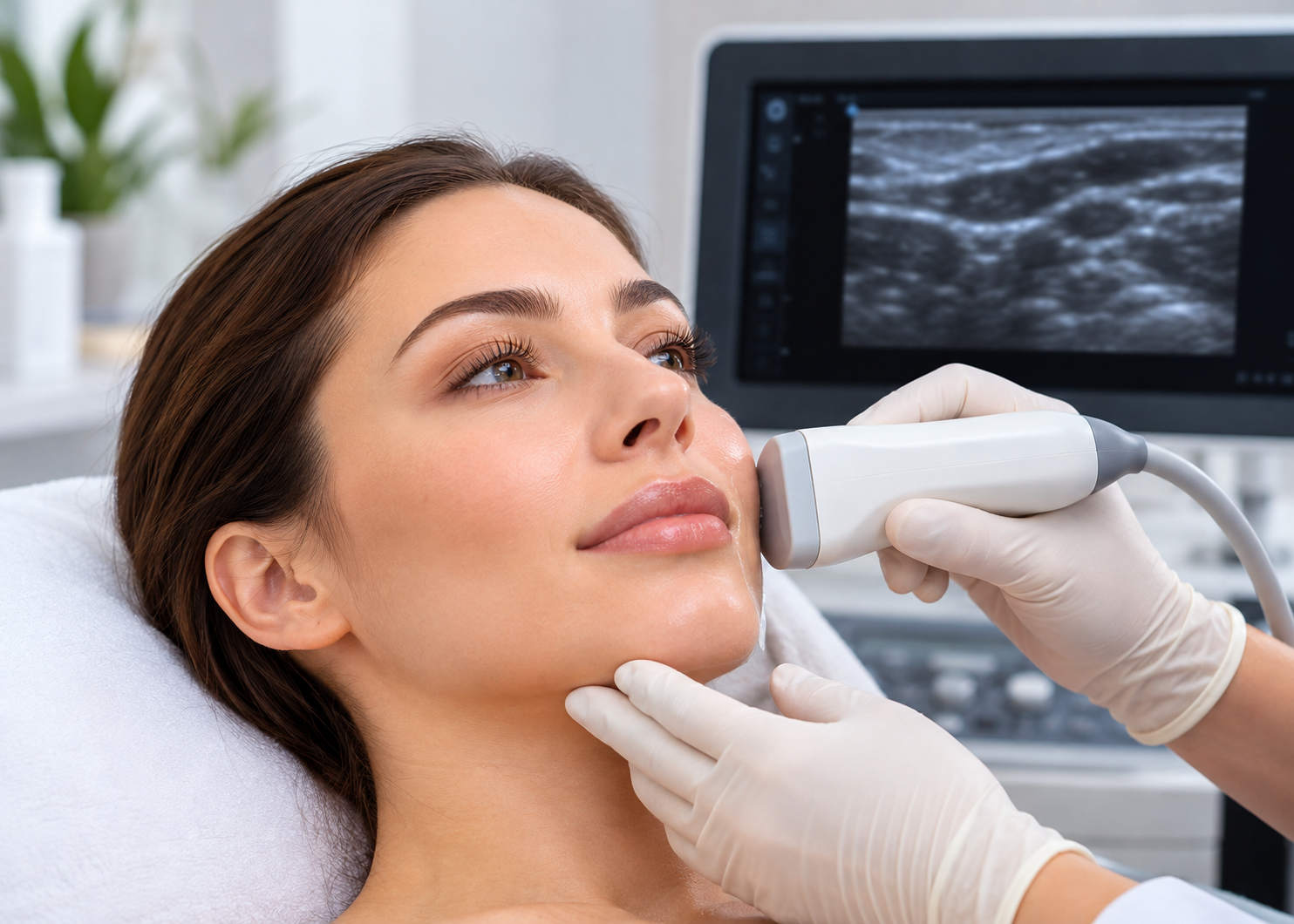
Filler migration is a condition in which injected dermal filler moves beyond its intended treatment area into nearby tissues. This may cause unwanted fullness, uneven contours, or changes in facial appearance. Common factors include injection technique, product volume, and individual anatomy. Ultrasound can help practitioners locate existing filler and assess its position. Although it cannot guarantee prevention, imaging supports more informed treatment decisions.

Professional guidance can help you understand your treatment options and plan your next steps. At Sadiya Clinic, you can discuss your concerns and explore suitable aesthetic treatments with a qualified provider. Concierge services offer added convenience for eligible treatments, subject to availability and applicable fees. Schedule a consultation today to discuss your needs and confirm suitable care options.

How Is Filler Migration Different From Overfilled Lips?

Filler migration occurs when injected material moves beyond its original placement, while overfilled lips result from excessive volume. Both conditions can change lip shape, but their underlying issues differ. Migration involves displacement into nearby tissues. Overfilling means too much product creates an enlarged appearance, even when the filler remains in its intended location.

A study in Dermatologic Surgery [1] used ultrasound to examine filler deposits across different tissue layers. Researchers identified migration in five of 27 patients with noninflammatory filler nodules. These findings show how imaging can reveal the location of injected material. However, the study did not directly compare migration with overfilled lips, so clinical assessment remains important when distinguishing these concerns.

What Causes Filler to Migrate?

Filler migration can result from excessive product volume, incorrect injection depth, poor placement, and repeated treatments. Facial movement and individual tissue structure may also affect filler distribution. In some cases, the product’s properties contribute to unwanted spread. However, migration does not occur in every patient, and several factors may influence its development.

Several factors may increase the likelihood of unwanted filler movement:

  • Excessive product volume: Too much filler can create pressure and unwanted fullness around the injection site.
  • Incorrect injection placement: Depositing filler in an unsuitable tissue layer may affect its distribution.
  • Repeated treatments: Adding more product before previous filler has sufficiently reduced can increase volume in the treated area.
  • Product characteristics: Different fillers vary in thickness, flexibility, and water absorption, which can influence their behavior within tissues.
  • Individual anatomy: Natural tissue structure and facial movement may affect how filler sits over time.

Although these factors can contribute to unwanted changes, filler migration is not always easy to identify from appearance alone. Swelling, overfilling, and other reactions may look similar. Therefore, a qualified practitioner should assess persistent changes before recommending treatment. Careful product selection, appropriate injection techniques, and follow-up can help support safer outcomes and reduce unnecessary corrective procedures.

What Are the Signs of Filler Migration?

Signs of filler migration include unexpected fullness beyond the treated area, blurred lip borders, uneven contours, and visible lumps. For example, lip filler may create a raised area above the upper lip. However, these changes can also result from swelling or excess product. A professional assessment can help identify the underlying cause.

Research published in Clinics in Dermatology [2] in 2024 examined ultrasound findings in nonvascular hyaluronic acid filler complications. The study highlights how different problems can produce similar visible changes. Therefore, unusual fullness, lumps, or uneven contours require careful assessment. Ultrasound may help practitioners identify underlying deposits and guide appropriate treatment decisions.

How Does Ultrasound Help Prevent Filler Migration?

Ultrasound helps reduce the risk of filler migration by showing practitioners where existing filler sits beneath the skin. This imaging method reveals tissue layers and nearby blood vessels. As a result, providers can assess previous injections and plan treatment more carefully. Although it cannot guarantee prevention, ultrasound supports better-informed decisions and more precise placement.

Identifies Existing Filler

High-frequency ultrasound can reveal filler deposits beneath the skin, including material that may not be visible externally. This helps practitioners assess previous injections before adding more product, reducing guesswork and supporting safer treatment planning.

Maps Facial Blood Vessels

Doppler ultrasound helps identify blood vessels near the planned injection site. This information supports careful needle placement and helps practitioners reduce vascular risks during treatment, especially in areas with complex facial anatomy.

Guides Precise Filler Placement

Real-time ultrasound can help practitioners observe the needle or cannula and the injected material. This allows better control over product placement and supports decisions based on the patient’s anatomy and existing filler distribution.

Supports Targeted Correction

When unwanted filler is identified, ultrasound can help locate the affected deposit. For suitable hyaluronic acid filler complications, practitioners may use imaging to guide hyaluronidase delivery toward the intended area.

Research published in the Journal of Cosmetic Dermatology [3], explored ultrasound-guided hyaluronic acid filler treatment. The study highlights how real-time imaging helps practitioners view tissue layers and nearby blood vessels. This guidance supports more precise injections and informed treatment planning. However, ultrasound cannot guarantee prevention of filler migration or eliminate every injection-related risk.

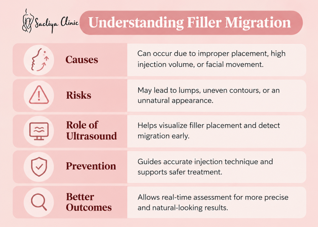
Filler migration explaining causes, risks, and how ultrasound helps assess filler placement and support safer treatment.

Can Filler Migration Be Corrected?

Yes, filler migration can often be corrected, depending on the product type, deposit location, and severity. A qualified practitioner first assesses the affected area before recommending treatment. Hyaluronic acid fillers may respond to hyaluronidase, an enzyme that breaks down the material. However, other fillers require different approaches, and some cases need careful monitoring.

Research published in the Journal of Cosmetic Dermatology [4] in 2024 examined how hyaluronidase affects different hyaluronic acid fillers. Using ultrasound, researchers measured changes in filler volume after treatment. The findings support dissolving as a potential option for unwanted HA deposits. However, treatment results vary, and professional assessment remains essential before choosing a corrective approach.

Book Your Filler Consultation at Sadiya Clinic

If you have noticed unusual fullness, uneven lip contours, or changes after filler injections, you may feel worried about your appearance. You might also feel unsure whether your filler has migrated or needs correction. Finding the right treatment can be confusing, especially when you want natural-looking results without unnecessary procedures.

At Sadiya Clinic, you can discuss your concerns and explore suitable options for managing filler migration. A personalized consultation can help clarify your needs and guide your next steps. Contact us today to learn more about available treatments and take the next step toward your aesthetic goals. Email us at sadiya@sadiyaclinic.com to get started.

FAQs

Can lip filler migrate years after injection?

Yes, lip filler can migrate years after injection, although other factors may cause similar changes. Hyaluronic acid filler can remain in tissues longer than expected. If you notice unusual fullness or changes in lip shape, consult a qualified practitioner. They can assess the area and recommend suitable treatment options.

Can migrated lip filler go away on its own?

Yes, migrated lip filler may fade naturally over time, depending on the product and its location. Hyaluronic acid filler gradually breaks down. However, unwanted fullness can persist for months or longer. Consult a qualified practitioner to assess the area and discuss suitable treatment options.

Can I prevent filler migration without dissolving my filler?

Yes, you may manage some filler concerns without dissolving the product. Avoid unnecessary top-ups and allow swelling to settle before considering further treatment. Choose an experienced injector who understands facial anatomy. If you suspect migration, seek professional assessment before deciding on your next step.

How long should I wait before getting more lip filler?

You should generally wait at least two weeks before considering additional lip filler. This allows swelling to settle and results to become clearer. However, timing varies by individual. Consult a qualified injector before booking another treatment, especially if you notice persistent lumps, unevenness, or unusual fullness.

Where can I get ultrasound-guided filler treatment in Northern Virginia?

You can find ultrasound-guided filler treatment at qualified aesthetic clinics in Northern Virginia. Contact a provider to confirm availability and practitioner training. Ask whether ultrasound is used during injection or only for assessment. Discuss your treatment history and goals before booking an appointment. This helps you choose suitable care for your needs.

About the Author

Dr. Aimen Arij, Pharm.D.

Dr. Aimen Arij, Pharm.D., is a licensed Clinical Pharmacist with over 6 years of experience in clinical and pharmaceutical practice. She has served as a Medical Science Liaison Executive for a multinational pharmaceutical company and has extensive experience working with international telepharmacy services. Her professional interests include medication safety, patient education, and translating clinical research into accessible, evidence-based health content.

Medically Reviewed By

Dr. Sadiya Naseem, DNP, FNP-BC

Dr. Sadiya Naseem, DNP, FNP-BC, is a board-certified Family Nurse Practitioner and the founder of Sadiya Clinic in Vienna, VA. She is a national injector trainer known for her ultrasound-guided approach to aesthetic treatments and her focus on safe, natural-looking results.

References

  1. Schelke, L. W., Decates, T. S., Cartier, H., Cotofana, S., & Velthuis, P. J. (2023). Investigating the anatomic location of soft tissue fillers in noninflammatory nodule formation: An ultrasound-imaging-based analysis. Dermatologic Surgery, 49(6), 588–595. 
  2. Gonzalez, C., Duque-Clavijo, V., Kroumpouzos, G., Garcia Concha, A. M., & Olmos Perez, M. (2024). Ultrasonographic features of nonvascular complications of hyaluronic acid fillers: A retrospective study at a reference center for dermatologic ultrasonography. Clinics in Dermatology, 42(5), 538–546. 
  3. Jung, J. Y., Choi, H., Han, S. C., Wanitphakdeedecha, R., Luo, Q., Zhou, L., No, J. I., Jang, W. E., & Kim, H.-J. (2025). Recommendations on ultrasound-guided hyaluronic acid soft tissue augmentation of the upper face in Asians. Journal of Cosmetic Dermatology, 24(1), Article e16759. 
  4. Bravo, B. S. F., Cavalcante, T., Silveira, C., Bravo, L. G., Zafra, M. C., & Elias, M. C. (2024). Resolve and dissolve—An ultrasound-guided investigation on the effects of hyaluronidase on different soft tissue fillers. Journal of Cosmetic Dermatology, 23(10), 3173–3181.

dermatologic surgery, Filler Migration, Overfilled lips, Sadiya Clinic, signs of filler migration, ultrasound, ultrasound and filler